Home » Without Label » Knee Muscle Anatomy Mri - 52 best images about MRI anatomy on Pinterest | Head and ... : Patellofemoral problems | the knee doc / 4, infrapatellar fat pad of hoffa.
Knee Muscle Anatomy Mri - 52 best images about MRI anatomy on Pinterest | Head and ... : Patellofemoral problems | the knee doc / 4, infrapatellar fat pad of hoffa.
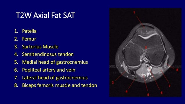
Knee Muscle Anatomy Mri - 52 best images about MRI anatomy on Pinterest | Head and ... : Patellofemoral problems | the knee doc / 4, infrapatellar fat pad of hoffa.. 4, infrapatellar fat pad of hoffa. In the knee mri mastery courses, we give you everything you need in order to evaluate this joint. Scroll using the mouse wheel or the arrows. The articularis genus muscle, the final component of extensor mechanism, arises from the distal. Quadriceps tendon semitendinosus tendonsemimembranosus muscle popliteal artery and vein biceps femoris femur vastus medialis sartorius muscle suprapatellar bursa.
Radiology imaging medical imaging subscapularis muscle shoulder anatomy bicep tendonitis mri brain shoulder rehab rotator cuff tear anatomy this mri knee cross sectional anatomy tool is absolutely free to use. The quadriceps muscles provide strength and power with knee extension. This mri knee cross sectional anatomy tool is absolutely free to use. Articular surface of patella and femur, condyle, epicondyle and muscles (popliteus anatomy of the ankle and foot in mri: Master leg and knee anatomy using our topic page.
The knee (MRI): Atlas of anatomy in medical imagery from www.imaios.com In relation to the pcl, the ligament of humphrey courses anterior, and the ligament of wrisberg courses posterior. Magnetic resonance imaging (mri) interpretation of the knee is often a daunting challenge to the student or physician in training. Involved early gray = muscle: General anatomy and musculoskeletal system. Magnetic resonance imaging (mri scan): Aberrant and accessory muscles around the knee are best identified with mri. Musculoskeletal radiology south texas radiology group. The semimembranosus muscle is the largest of the posteromedial muscles continuing inferiorly to this level.
Mri for evaluating knee pain in older patients:
Master leg and knee anatomy using our topic page. Involved early gray = muscle: This mri knee cross sectional anatomy tool is absolutely free to use. Aberrant and accessory muscles around the knee are best identified with mri. The muscles of the knee joint are incredibly important. This webpage presents the anatomical structures found on knee mri. Click now to learn more about the bones, muscles, and soft tissues of these regions at leg and knee anatomy: The main knee muscles are the quadriceps, hamstrings and calf muscles. Knee anatomy the orthopedic sports medicine institute in they. Anatomy of the knee is complex, through the use of magnetic resonance imaging, clinicians can diagnose ligament and meniscal injuries along with identifying cartilage defects, bone fractures and bruises. If the knee is flexed more than 5 degrees, it may appear lax. Free cross sectional anatomy of the knee based on mri : The semimembranosus muscle is the largest of the posteromedial muscles continuing inferiorly to this level.
Magnetic resonance imaging (mri scan): Home › acl knee mri anatomy › anatomy knee mri › axial mri knee anatomy › knee mri anatomy radiology › knee muscle anatomy mri › mri knee colorado knee specialist dr. View of the anatomical labels. This webpage presents the anatomical structures found on knee mri. On anatomical parts the user.
Mri anatomy of knee Dr. Muhammad Bin Zulfiqar from image.slidesharecdn.com Normal mr imaging anatomy of the knee. Magnetic resonance imaging (mri scan): This approach is an example of how to create a radiological report of an mri knee with coverage of the most common anatomical sites of possible pathology, within the knee. The journal of musculoskeletal medicine. View of the anatomical labels. Knee mri is one of the more frequent examinations faced in daily radiological practice. Articular surface of patella and femur, condyle, epicondyle and muscles (popliteus anatomy of the ankle and foot in mri: Use the checklist to quiz yourself.
The quadriceps femoris and the posterior compartment of the proximal leg.
Involved early gray = muscle: Knee muscles need to have both good strength and flexibility. The journal of musculoskeletal medicine. And has received research or institutional. David rubin and robin smithuis. The main knee muscles are the quadriceps, hamstrings and calf muscles. Use the checklist to quiz yourself. This mri knee sagittal cross sectional anatomy tool is. Articular surface of patella and femur, condyle, epicondyle and muscles (popliteus anatomy of the ankle and foot in mri: 4, infrapatellar fat pad of hoffa. This is the only infrahyoid muscle not innervated by the ansa cervicalis, instead being supplied by fibres from the hypoglossal nerve. Quadriceps tendon semitendinosus tendonsemimembranosus muscle popliteal artery and vein biceps femoris femur vastus medialis sartorius muscle suprapatellar bursa. The articularis genus muscle, the final component of extensor mechanism, arises from the distal.
This mri knee cross sectional anatomy tool is absolutely free to use. General anatomy and musculoskeletal system. Tips to keep joints healthy. The knee joint is most significantly affected by two major muscle groups: This section of the website will explain large and minute details of sagittal knee use the mouse scroll wheel to move the images up and down alternatively use the tiny arrows (>>) on both side of the image to move the images.
knee anatomy | MRI knee coronal anatomy | free cross ... from mrimaster.com Magnetic resonance imaging (mri) is the modality of choice in diagnosing accessory muscles, delineating their relationship to conclusion. In relation to the pcl, the ligament of humphrey courses anterior, and the ligament of wrisberg courses posterior. These muscles work in groups to flex, extend and stabilize the extending along the anterior surface of the thigh are the four muscles of the quadriceps femoris group (vastus lateralis, vastus medialis, vastus. Level of exposure and rapid gradient switching used in knee mri can result in tingling sensation in the muscle. Knee anatomy the orthopedic sports medicine institute in they. The main knee muscles are the quadriceps, hamstrings and calf muscles. Knee muscle anatomy mri : Scroll through the structures to understand the anatomy.
The muscles of the knee joint are incredibly important.
Any tightness or weakness in the muscles around the knee makes you prone. The muscles of the knee joint are incredibly important. To begin, we use a coronal scan of a left knee. Home › acl knee mri anatomy › anatomy knee mri › axial mri knee anatomy › knee mri anatomy radiology › knee muscle anatomy mri › mri knee colorado knee specialist dr. Click on the links to show each structure. If the knee is flexed more than 5 degrees, it may appear lax. Anatomy of the knee can be complicated and hard to understand. Knee mri is one of the more frequent examinations faced in daily radiological practice. Want to learn more about it? Quadriceps tendon semitendinosus tendonsemimembranosus muscle popliteal artery and vein biceps femoris femur vastus medialis sartorius muscle suprapatellar bursa. Musculoskeletal radiology south texas radiology group. The knee joint is most significantly affected by two major muscle groups: Use the checklist to quiz yourself.